This post is a simplified description of the various types of connective tissue that make up our bodies and where some of the connective tissue types are found. Connective tissue is also found in other parts of the body which are not mentioned here. This list has been condensed to focus on muscle anatomy and give you a general understanding of the anatomy involved.
Connective Tissue
Connective tissue is a term used to describe the cellular matrix (types of cells) that make up the different structures in our bodies which are listed below;
- Loose Connective Tissue
- Areolar Connective Tissue
- Found in different layers of skin, around blood vessels, nerves and organs
- Adipose Tissue
- Found around the heart, kidneys, padding around joints and behind the eyeball in the eye socket
- Reticular Connective Tissue
- Found in the supporting framework (stroma) of the liver, spleen, lymph nodes and around blood vessels and muscles
- Areolar Connective Tissue
- Dense Connective Tissue
- Dense Regular Connective Tissue
- Forms tendons, most ligaments and aponeurosis which are sheet-like tendons that attach muscle to muscle, or muscle to bone
- Dense Irregular Connective Tissue
- Found in fascia beneath the skin, around muscles & other organs including; the pericardium of the heart & heart valves and periosteum of bone
- Elastic Connective Tissue
- Found in lung tissue, walls of elastic arteries and vocal chords
- Dense Regular Connective Tissue
- Cartilage
- Hyaline Cartilage
- Found at the ends of long bones, nose and parts of your larynx
- Fibrocartilage
- Found between your intervertebral discs, menisci of the knee, pubic symphysis and portions of tendons that insert into cartilage
- Elastic Cartilage
- Found in part of your external ear and on your larynx (epiglottis)
- Hyaline Cartilage
- Bone Tissue
- Compact Bone
- Found in bone tissue
- Compact Bone
- Liquid Connective Tissue
- Blood
- Forms within blood vessels (arteries, arterioles, capillaries, venules, and veins) and in the heart chamber
- Blood
Muscles
Skeletal muscle is the (voluntary contractile tissue) type of muscle that moves your skeleton. It is composed of muscle cells (fibers), layers of connective tissue (fascia) and nerves and blood cells.
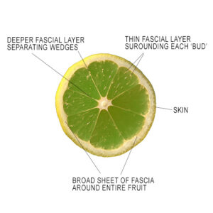
The infrastructure of a muscle is very similar to a tropical fruit like a lime. A broad sheet of fascia encases the whole fruit, deeper layers of fascia separate the fruit into ‘wedges’ and then a thin coating of tissue surrounds each individual, tiny bud of fruit. If we use this analogy then a layer of fascia (epimysium) encases the muscle belly, a deeper layer (perimysium) wraps the long muscle fibers into bundles called ‘fascicles’ and each microscopic muscle fiber is bound in fascia (endomysium). Unlike a fruit, a muscle’s layers of connective tissue merge at either end of the muscle to form a strong tendon. The tendon attaches the muscle to a bone. A fruit only attaches at one end to a branch via its stem.
Muscle tissue has three specific characteristics that help distinguish it from other body tissues.
- Skeletal muscle tissue has a striated texture – (Much like a plank of wood) This is different from tendons which have a much smoother feel. The fibrous texture of muscle tissue is caused by the muscle fiber bundles running in a particular direction.
- Muscle fiber directions – You can feel the direction of the muscle fiber to determine which muscle you are feeling. Depending on the shape and design of the muscle, the direction of the muscle could be parallel, diagonal or get closer together. A good example of this is your erector muscles v.s. your trapezius or rhomboids.
Your erector muscles run “vertically” up your back while your rhomboids run “horizontally” and your mid trapezius runs on more of an “oblique angle” than your rhomboids. - Muscle tissue can be relaxed or contracted – When a muscle is relaxed it feels more malleable. When it is contracted it is more solid. As the muscle tissue tension changes, the tendons and fascia also become more loose or taut.
Tendons
Simply put, tendons attach muscles to bone. …But what they actually do is connect muscles to the periosteum which is the connective tissue that surrounds the bone. Tendons are made of dense connective tissue shaped into bundles of parallel collagen fibers. Each end of a muscle has one or more tendons.
Tendons also come in a variety of shapes and sizes. Some are short and wide. Others are long and thin. There is also something called an aponeurosis which is a broad flat tendon. All tendons have a tough, smooth feel to them.
Ligaments
Ligaments connect bones together at a joint. They strengthen and stabilize joints. Ligaments are also made of very dense connective tissue. Ligament fibers have an uneven configuration whereas a tendon has a parallel fiber arrangement. Ie Deltoid ligament in the ankle and foot.
Some ligaments cross a joint and blend in with the joint capsule while others can span across several bones. (Ie Supraspinous ligament which extends inferiorly from the ligamentum nuchae down the spine as it attaches to each spinous process of the thoracic and lumbar vertebrae.
The ligamentum nuchae is a sheet of connective tissue that looks like a fin which runs along the sagittal plane from the external occipital protuberance to the spinous process of C7 in the neck. Its job is to help stabilize the head and neck as well as being an attachment site for muscles.
Ligaments sometimes have a taut, dense feel to them and sometimes you can feel their fiber directions. In order to distinguish a ligament from a tendon feel the tension and attachments. A tendon connects muscle belly to a bone, while a ligament attaches a bone to a bone. A tendon can become taut or slack whereas a ligament will remain taut throughout the entire action of a muscle contraction.
Fascia
Fascia is a type of dense connective tissue, like ligaments and tendons. It is a continuous sheet of membrane located under the skin and around muscles and organs. It is a system of tissue that forms a three-dimensional matrix extending from head to toe throughout the entire body.
There are 2 types of fascia:
- Superficial Fascia – Superficial fascia is located immediately deep to the skin and covers the entire body. While many people think of it as being thin, it can vary in thickness. Examples of this would be the thin layer on the back of a hand and the very thick layer on the sole of your foot.
- Deep Fascia – Deep fascia surrounds muscle bellies, holding them together and separating them into functional groups. Ie. various compartments of the forearm. It also fills in the spaces between muscles as well as carries blood vessels and nerves. Portions of the deep fascia penetrate into the muscle belly and encase each tiny muscle fiber. It can be hard to feel because it is so spread out.
Retinaculum
A retinaculum is a structure that holds an organ or tissue in place. It is a transverse thickening of deep fascia which straps tendons down in a particular location or position. Ie the superficial layer of the carpal tunnel which holds the flexor tendons and nerves in place.
Aponeurosis
An aponeurosis is a flat sheet of tendon-like tissue on the surface of some muscles that function as insertion sites for muscle fascicles that anchors the muscle or connects it with the part that the muscle moves.
Interosseous Membrane
Interosseous membrane is a fibrous tissue that binds the radius to the ulna, ensuring they remain a fixed distance apart. Tension changes occur in the interosseous membrane during movements of the forearm.
The interosseous membrane of the leg is also referred to as the middle tibiofibular ligament. This ligament extends through the fibula and tibia’s interosseous crests and separates the muscles in the back of the leg from the muscles located in the front of the leg.